EDC Impressum & DSE

Electrophoresis
Development & Consulting
Dr. Hanspeter Schickle
Wolfgang Gstrein

Datenschutzerklärung >>

Coomassie-Violet Staining

Lipohilic proteins will disappear or at least show broader bands during the staining procedure with alcohol containing recipes. As a non-alcoholic procedure we suggest to use the “semi-colloidal Coomassie-Violet” staining.
Coomassie-Violet R200 (Acid Violet; CI 42650) Sigma #210579-50g

The procedure: all steps in ambient temperature!

April 28, 2023

Fixing

20% TCA

200 ml

30 min

Rinsing 1

10% Ethanol

200 ml

20 min

Rinsing 2

H2O dist.

200 ml

20 min

Staining

0.01% Coomassie-Violet
3,4% (w/v) Phosphoric acid + 3% (v/v) HAc
Mix shortly before use!

400 ml

30 - 50 min
(staining begins within minutes)

Enhancing
(if necessary)

200 ml H2O
+ 3 ml dye concentrat (= 0.0015% violet)
+ 3 ml hot coomassie concentrate (= 0.00045% R350)

200 ml

overnight

Washing + Preserving

5% Ethanol + 5% Glycerin + 5% Ethylenglycol

200 ml

20 min

Stock solutions:
Dye-Concentrate (10 x conc): 0,1% Coom.Violet = 1 g + 1 l H2O dist

Mixing of 400 ml staining solution: To be done shortly before use!
ca. 340 ml H2O dist. + 40 ml dye-conc. + 12 ml HAc + 17 ml H3PO4

Abbrev.: TCA = Trichloric Acide, HAc = Acetic acid

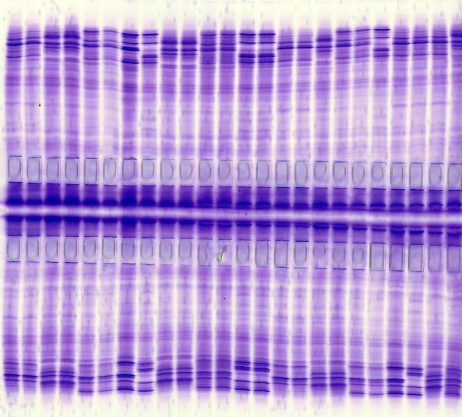
half IEFGel bidirect 104S stained with Coomassie-Violet incl. Enhancing

IEFGel bidirect cereal EQ-type

EDC Electrophoresis Development & Consulting, Vor dem Kreuzberg 17, 72070 Tübingen (Germany)