|
Gels polymerized on fluorescent film-supports (Gelbond films, Polyester, should be visualized with dyes that can be excited between 440 nm and 500 nm, or higher nm.
For visualization of DNA-fragments:
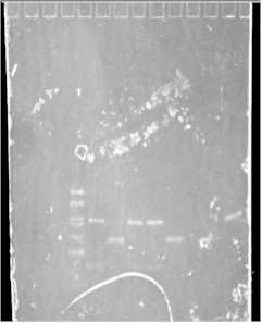
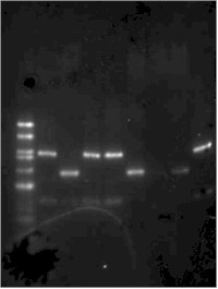
Camprex's GelStar and Sybr Green I and II are such molecules. Under this condition the plastic foil shows no self-fluorescence, figure 2. GelStar and Sybr Green I can be applied to the double-stranded DNA (dsDNA) before the run (tagging) and will stay at the double strand during the whole electrophoretic run without changing the fragment's mobility. After the electrophoretic procedure the gel can be taken to the imaging process directly.
Single stranded DNA or RNA can be visualized with Sybr Green II by also tagging the samples or of course equilibrating the gel after the run in the staining solution.
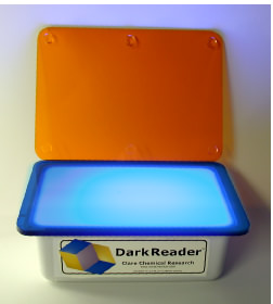
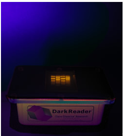
As visualization hardware a socalled blue light table should be used, see figure 5.
The sensitivity of these stains is about 500 pg (dsDNA), 2 ng (RNA, ssDNA) and 2 ng (proteins).
Silver-stained DNA-fragments and proteins: 2 pg.
Fluorescence dyes with exciting wavelengths between 440 and 500 nm
|

