|
The picture on the left side shows a 2D-separation of serum proteins.
Beside of the IgGs only 3 other proteins can be found between the pH-intervall of interest (6-11):
1. The hemolysate proteins (hemoglobins ) at pH 7-7.2 (16 000)
(It is important to identify the Hemolysate-proteins in a general visualization procedure: A special PAG-gel pH 6-11 has small slots at the edges to pipet diluted hemolytate onto the gel. Hemolysate proteins in the serum or the CSF-lanes can now be tagged)
2. The Prostaglandin D Synth. (ß-trace) at pH 6.8 (25 000)
3. The Cystatin C (g-trace) at pH 11 (35 000),
(This protein can only be found in th CSF and is located outside of the IgG region. No mismatch analysis should be possible), a degradation product is at pH 8 (30 000).
These are the reasons why:
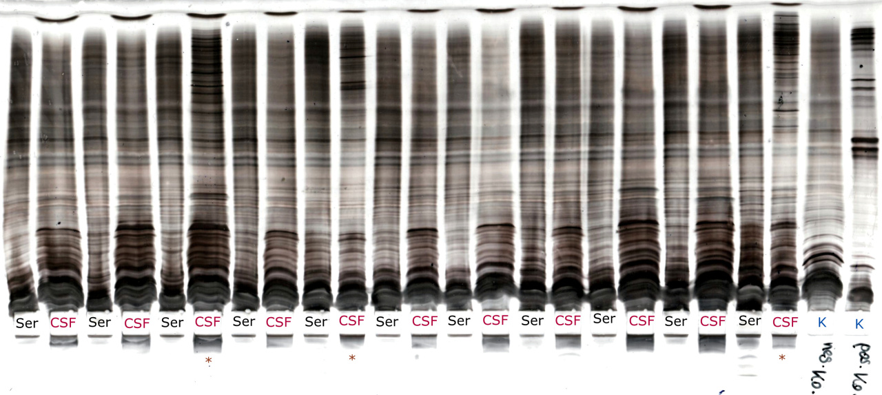
If a general visualization is chosen the amount of bands in the CSF should be more than 2-3 to be typed as positiv.
|

